The brain's rhythms in plain words
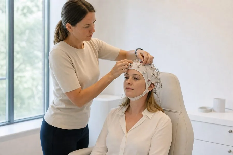
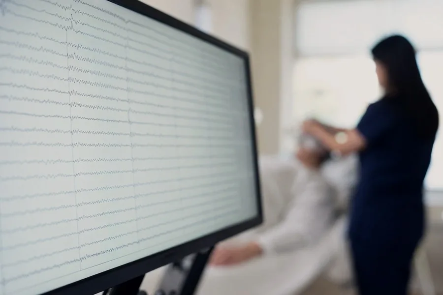
Brain cells communicate with tiny electrical signals. When large numbers of them work together, the combined activity forms waves that can be measured through the skull. An EEG trace shows these waves as lines across a screen, one for each pair of electrodes, and they are described by how fast they rise and fall.
When you sit relaxed with your eyes closed, a steady rhythm called alpha usually appears over the back of the head. It fades when you open your eyes or start concentrating, and faster activity takes over. As you become drowsy and fall asleep the waves slow down, which is normal. A neurophysiologist reads the trace by checking that these patterns appear where and when they should.
What a normal EEG looks like — and why it still helps
A normal EEG means the background rhythms are appropriate for your age, similar on both sides of the head, and respond as expected to opening the eyes, deep breathing and flashing light, with no worrying bursts during the recording.
Normal results are common, including in some people who go on to be diagnosed with epilepsy, because a recording samples a short period. That does not make the test wasted. It can make some conditions less likely, help a doctor decide whether more tests are needed, and provide a baseline to compare with later. It does not mean your symptoms are imagined.
What 'abnormal' can mean
Abnormal brain activity on an EEG covers a range of findings, and the word on its own does not name a cause. Slowing means waves slower than expected for the situation. It can be widespread, as can happen with drowsiness, some medicines, after a seizure or when the body's chemistry is upset, or limited to one area, which may point to a local problem worth investigating further.
Spikes and sharp waves are brief, pointed changes that stand out from the background. They are called epileptiform because they are associated with a tendency to seizures. A photoparoxysmal response is a burst of this kind triggered by the flashing light, and suggests sensitivity to flicker.
Some harmless variants look unusual to an untrained eye but are recognised as normal. Telling these apart from meaningful findings is the reason an EEG is read by a specialist.
What an EEG is used to investigate
Lists such as '10 conditions diagnosed with an EEG' are common online, but an EEG rarely diagnoses anything on its own. It is one piece of information, used alongside history, examination and other tests. Doctors may request one when looking into the following.
What an EEG cannot show
An EEG measures function, not structure. It cannot show a tumour, a stroke, multiple sclerosis or bleeding inside the head; those need a scan, usually MRI. A normal EEG therefore does not mean the brain has been checked for every problem.
It also does not measure intelligence, personality or mental health, and it cannot read thoughts. It is not usually helpful for investigating headaches on their own, and your GP or specialist will consider whether a scan or other assessment is more appropriate.
How EEG results are reported and explained
At Bridge House Clinic, Dr Rodrigues analyses the full recording and writes the report. It usually starts with a description of what was seen, such as the background rhythm and the response to deep breathing and flashing light, then gives an interpretation of what those findings may mean in relation to the question asked. The report goes to you and, with consent, to your referring clinician. The clinic has no neurologist, so the doctor looking after you explains how the EEG results fit with everything else.
This article is general information and not a diagnosis. If you are concerned about blackouts, seizures or confusion, see your GP or call NHS 111. Call 999 if a seizure lasts more than five minutes or keeps repeating, or for sudden weakness or numbness of the face, arm or leg, slurred speech or new confusion, which may be a stroke.
Common questions
Can an EEG show a brain tumour? +
No. An EEG records electrical activity, not structure. Sometimes it shows slowing in one area that prompts a scan, but only imaging such as MRI can show a tumour.
Can an EEG show anxiety or depression? +
No. It does not assess mental health, mood or personality. It is used to look at patterns of brain activity linked mainly to seizures and changes in consciousness.
What does slowing on an EEG mean? +
It means some waves are slower than expected. This has many possible causes, from drowsiness and medicines to recovery after a seizure or a local problem, so it is always interpreted in context.
Is a normal EEG a good result? +
It is reassuring and useful, but it does not rule everything out. Your doctor will consider it together with your history and any other tests.
Who explains my EEG results? +
Dr Rodrigues writes the report, and your referring doctor or specialist explains what it means for you and decides on next steps.